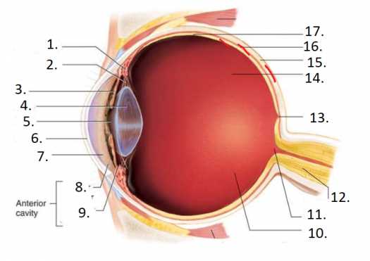
The intricate structure responsible for our sense of sight is a marvel of biological engineering. This complex system enables us to perceive the world in vivid detail and color, transforming light into meaningful images. Each component plays a crucial role in this remarkable process, contributing to our ability to navigate our surroundings.
In this exploration, we will delve into the various elements that come together to create our visual experience. From the outer layers that protect and guide light, to the inner workings that interpret signals, understanding these components will enhance our appreciation of how we see. The synergy among these structures is essential for achieving the ultimate clarity in vision.
By examining each section individually, we can gain insight into their specific functions and interactions. This knowledge not only illuminates the complexity of the visual system but also highlights the importance of each element in maintaining our ability to perceive the world around us.
Understanding the Eye Structure
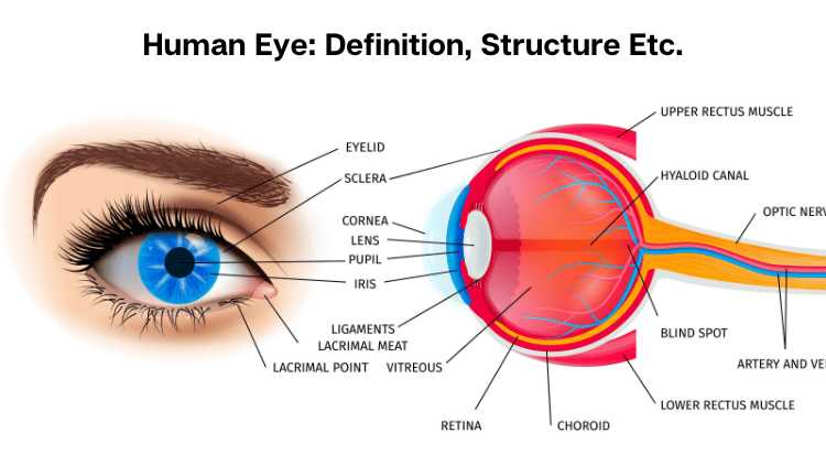
The human visual organ is a complex and fascinating system, designed to capture and process light, enabling the perception of the surrounding world. Each component plays a vital role, working in harmony to translate visual stimuli into signals interpreted by the brain. This intricate design reflects both the evolutionary advancements and the remarkable adaptability of sensory mechanisms.
Anatomically, this sensory organ can be divided into several regions, each with distinct functions. From the outer layers that protect and maintain integrity to the inner structures responsible for processing images, each element contributes to overall functionality. Understanding how these components interact enhances our appreciation of this remarkable feature of biology.
The intricate relationships between these structures facilitate processes such as focusing, adjusting to light variations, and transmitting information. The coordination of muscles and the biochemical processes involved are equally essential, highlighting the sophistication of the system. By exploring these elements, one can gain insight into not only how vision works but also how crucial it is to daily life.
Basic Anatomy of the Eye
The intricate structure of vision involves various components that work harmoniously to facilitate sight. Understanding this framework is essential for appreciating how we perceive the world around us.
Key elements include:
- Cornea: The transparent outer layer that refracts light.
- Iris: The colored part that controls the size of the pupil.
- Pupil: The opening that regulates light entry.
- Lens: The flexible structure that focuses light onto the retina.
- Retina: The light-sensitive layer that converts images into neural signals.
- Optic nerve: The pathway transmitting visual information to the brain.
Each of these components plays a vital role in the overall functioning of vision, ensuring clarity and detail in what we observe.
Function of the Cornea
The transparent, curved surface of the visual system plays a vital role in directing and focusing light toward the retina. Its ability to bend and filter incoming rays ensures that the image seen is clear and sharp. Without proper function, visual clarity would be compromised, affecting how one perceives the world.
Light Refraction and Focusing
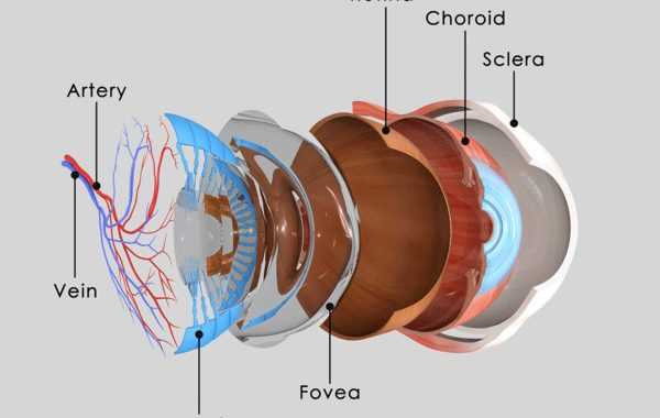
The cornea is responsible for bending light as it enters, helping it to reach the focal point of the visual sensory organ. This process is essential for producing accurate images, especially for objects that are both near and far.
- Refraction allows light to enter at the correct angle.
- Helps focus light before it passes through the lens.
- Ensures the clarity of images and visual details.
Protection and Filtering
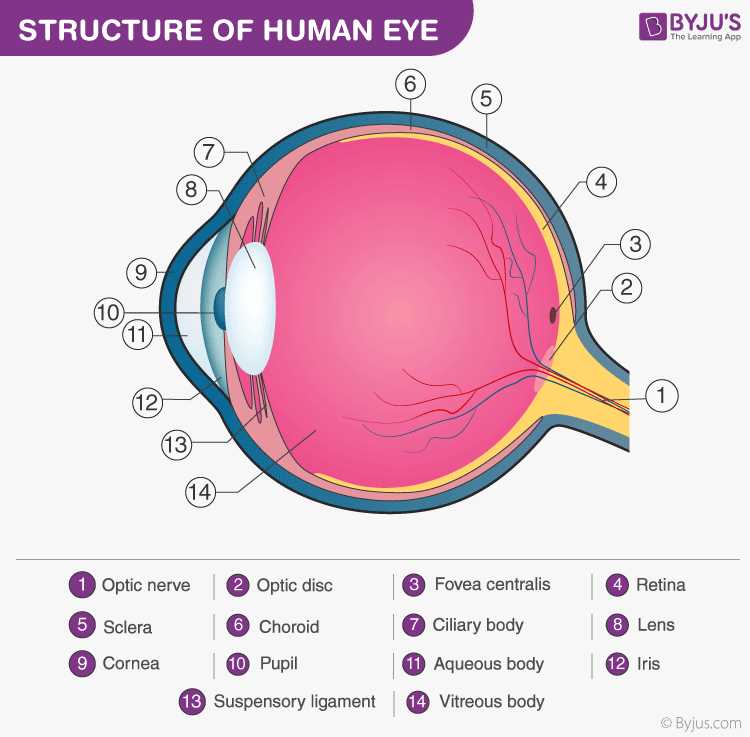
Beyond its optical functions, the outermost layer offers protection by acting as a barrier against harmful particles, dust, and microorganisms. Additionally, it helps filter out some ultraviolet (UV) light, reducing the potential for damage to the deeper structures.
- Prevents foreign objects and pathogens from reaching sensitive tissues.
- Blocks excessive UV rays to minimize long-term damage.
- Assists in maintaining a healthy environment for proper vision function.
The Role of the Lens
The lens is a crucial component responsible for focusing incoming light onto the sensitive surface within the visual system. Its primary function is to adjust the focus of light, ensuring that images are sharp and clear. This adjustment allows the system to perceive objects at different distances with precision.
The lens accomplishes this by changing its shape, which is known as accommodation. This process enables the system to focus on objects both near and far by altering the curvature of the lens to bend the light rays appropriately.
- Focusing light: The lens bends light rays to create a sharp image on the receptive layer, allowing clear vision.
- Adapting to distance: By changing shape, the lens allows the system to focus on objects at varying distances.
- Maintaining clarity: The lens ensures that images remain sharp by precisely managing light exposure.
In this way, the lens plays a vital role in maintaining clear vision, ensuring that the details of the world are properly captured and processed by the visual system. Without proper lens function, focusing errors or blurry images would result, making it impossible to perceive the environment accurately.
Importance of the Retina
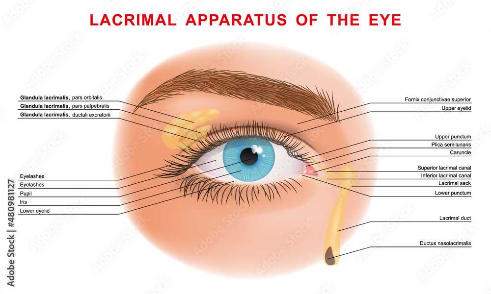
The retina is a vital component responsible for capturing and processing visual information. Its role is essential in translating light signals into electrical impulses that the brain can interpret, enabling us to perceive the world around us. Without proper functioning of this structure, the ability to see and interpret visual stimuli would be compromised, leading to impaired vision or even blindness.
Located at the back of the optical organ, this layer is composed of highly sensitive cells that react to light. These cells, known as photoreceptors, convert the light entering the pupil into signals that are then sent to the brain through the optic nerve. This process is crucial for creating the images we perceive, allowing us to navigate and interact with our environment effectively.
| Feature | Function |
|---|---|
| Photoreceptors | Convert light into electrical signals |
| Macula | Responsible for sharp central vision |
| Optic Nerve | Transmits visual information to the brain |
Maintaining the health of this structure is crucial for long-term vision. Any damage or disease affecting it, such as macular degeneration or diabetic retinopathy, can lead to serious visual impairments. Therefore, understanding its role emphasizes the importance of regular check-ups and protective measures to preserve vision quality throughout life.
How the Iris Controls Light
The ability of the human visual system to adapt to varying light conditions is a remarkable feature. This adjustment is essential for maintaining clear and sharp vision under different environmental lighting scenarios. One key mechanism responsible for controlling the amount of light entering the vision organ is a specialized structure that regulates the size of the opening through which light passes.
Regulating Light Entry
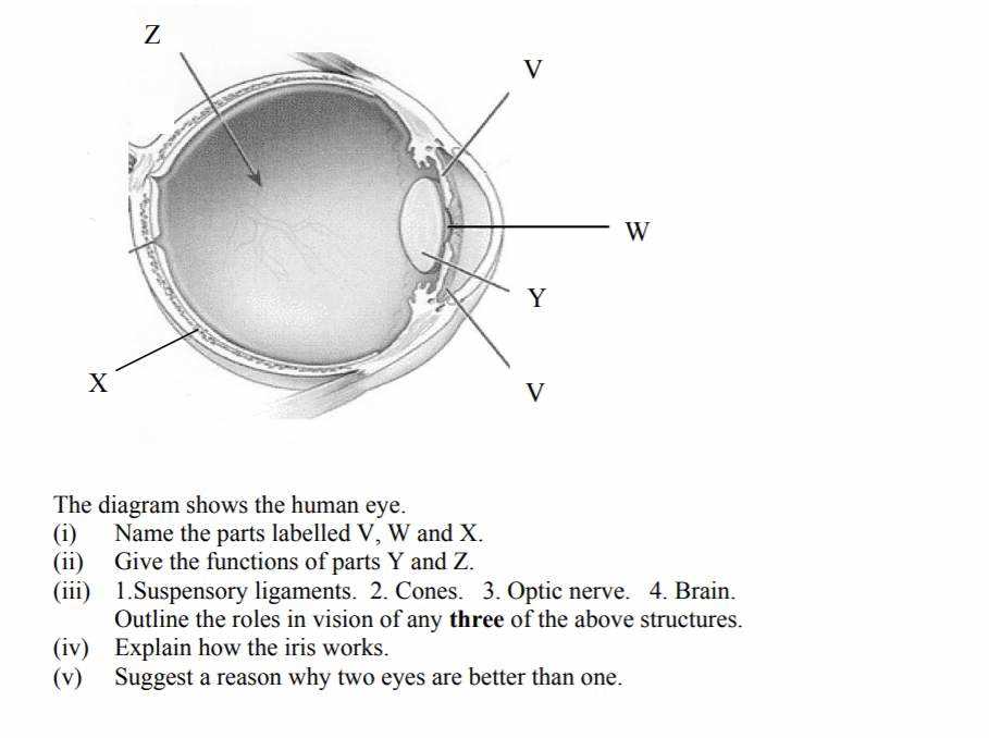
The primary function of this structure is to manage the intensity of light entering the sensitive layers where visual information is processed. By changing the size of the central aperture, it allows more or less light to reach the internal surface, optimizing vision in both bright and dim conditions.
- In bright light, the aperture constricts, reducing the amount of incoming light to prevent overload and discomfort.
- In low-light conditions, the aperture dilates, allowing more light to enter and improve visibility.
Automatic Adjustment
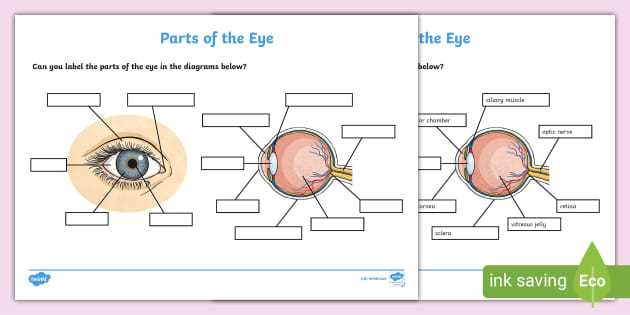
This adaptation occurs almost instantly, with the surrounding muscles responding quickly to changes in lighting. Such reflexes help to maintain optimal vision without conscious effort, ensuring that the sensory cells are never overwhelmed or under-stimulated.
Understanding the Pupil’s Mechanism
The mechanism that controls the entry of light into the visual system is an essential function of the organism. This process, occurring in a specialized area of the visual structure, adjusts to varying lighting conditions to ensure proper image perception. The system’s sensitivity to different levels of brightness is key to optimizing vision, providing clarity in both bright and dim environments.
How It Responds to Light
The control system responds dynamically to changes in light intensity. This process involves rapid contraction and dilation, altering the amount of light that passes through. This adjustment ensures the optimal amount of illumination for proper focus and clarity. When bright light is detected, the structure contracts, limiting exposure. In low-light conditions, it dilates to allow more light in, improving vision in darker settings.
Key Factors Affecting the Mechanism
- Brightness: Light levels directly influence the size adjustments, with brighter conditions triggering constriction and dimmer settings causing dilation.
- Emotional and Physiological States: Changes in mood or physical reactions can also cause temporary alterations in its size.
- Age: As organisms age, the ability to adjust may slow down, impacting visual adaptation to various environments.
The Function of the Sclera
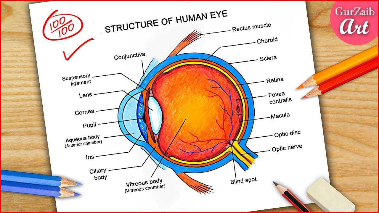
The sclera plays a critical role in maintaining the structural integrity and overall function of the visual organ. It serves as the outermost protective layer, safeguarding the delicate components inside from potential damage. By offering support and stability, the sclera also contributes to the proper alignment and functioning of all surrounding tissues.
Structure and Protection
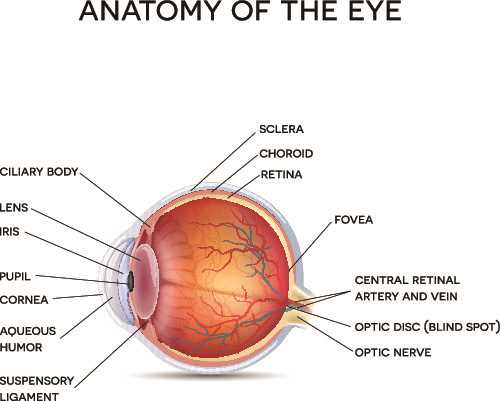
This tough, fibrous tissue provides a rigid framework that prevents excessive deformation of the entire organ. It shields the inner elements from physical impact and pressure, allowing them to work harmoniously. The sclera is also the attachment site for the muscles that control movement, ensuring precise and coordinated motion.
Connection to Other Structures
In addition to its protective role, the sclera works closely with other components to facilitate communication and interaction within the complex system. It helps maintain the proper position of the components, contributing to the efficiency of visual processing.
| Feature | Function |
|---|---|
| Protective Barrier | Prevents damage and safeguards internal tissues |
| Structural Support | Maintains the shape and alignment of the visual system |
| Attachment Point | Serves as the anchoring site for muscles controlling movement |
How Vitreous Humor Affects Vision
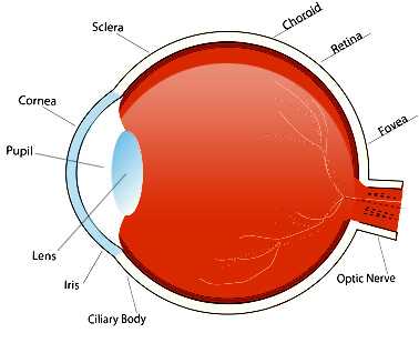
The clear, gel-like substance found in the central region of the visual organ plays a significant role in shaping the way we perceive the world around us. It fills the space between the lens and the retina, contributing to the overall functionality of the visual system. Its consistency and transparency are essential for maintaining sharpness and clarity of what we see.
The Role of Vitreous Humor in Visual Clarity
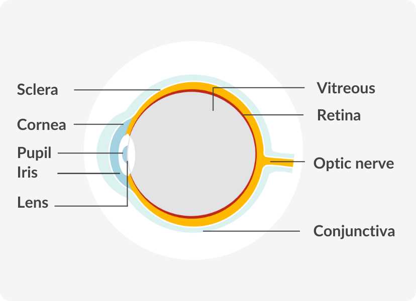
Vitreous humor serves as a cushion, helping to maintain the spherical structure of the visual system and protect sensitive components from pressure or damage. Its transparency is crucial in allowing light to pass through unobstructed, ensuring that images reach the retina clearly and without distortion.
- Maintains the shape of the visual structure.
- Ensures the proper transmission of light.
- Helps prevent physical damage from external forces.
How Changes in Vitreous Humor Impact Vision
Over time, the consistency of this gel-like substance may change, leading to various visual disturbances. As it begins to liquefy or develop opacities, it can cause symptoms such as floaters, blurring, or even vision loss in severe cases. These changes can interfere with the transmission of light and affect the clarity of images on the retina.
- Floaters: Small, dark specks or strings that appear in the field of vision.
- Blurred vision: Decreased sharpness due to uneven distribution or density of the substance.
- Potential retinal damage: If the vitreous pulls away from the retina, it could lead to more severe complications.
Significance of the Optic Nerve
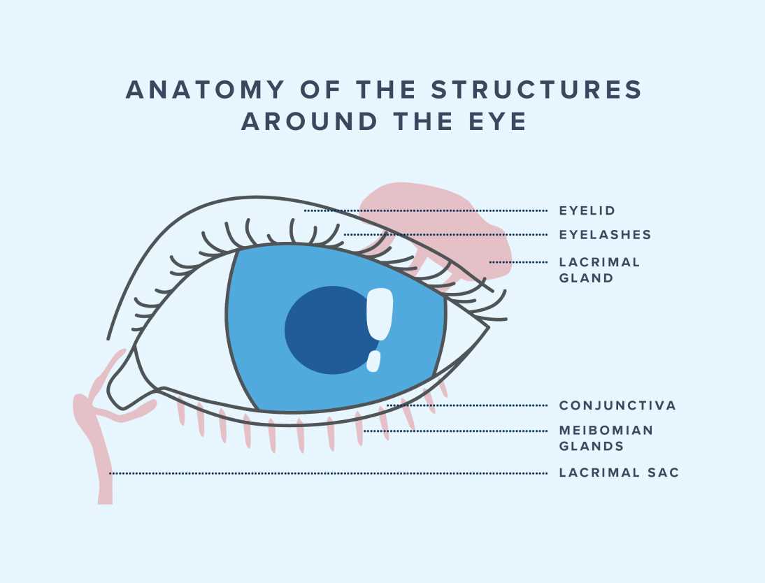
The optic nerve plays a crucial role in the process of visual perception, serving as the primary pathway for transmitting information from the light-sensitive cells to the brain. This bundle of fibers allows the brain to interpret the visual signals received, effectively translating what we see into meaningful images.
Role in Visual Information Processing
Once light is captured and converted into electrical signals by the retina, the optic nerve transmits these signals to the brain, specifically to the visual cortex. This is where complex processing occurs, allowing us to recognize shapes, colors, and movement. Without the optic nerve, visual communication between the eyes and the brain would be impossible.
Impact on Vision Loss
A disruption or damage to the optic nerve can lead to partial or complete loss of sight. Conditions such as glaucoma, optic neuropathy, and other related disorders can impair its function, affecting the transmission of visual data and ultimately leading to impaired perception of the surrounding environment. Protecting the health of the optic nerve is essential for maintaining clear and accurate vision throughout life.
Connection Between Eye Parts
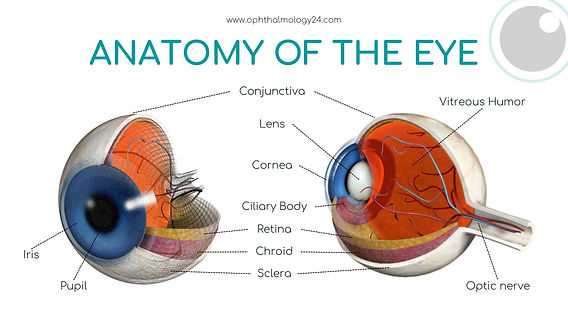
The different components of visual systems work in harmony to create a clear and accurate representation of the transmitted signal. Each section plays a crucial role in the overall process, where the interaction between them ensures that the data received is correctly interpreted. These elements communicate through precise synchronization, allowing for the proper analysis of the information.
Signal transmission begins with the initial reception of light waves, which are then processed and converted into digital form by specialized receptors. These digital signals are passed through a series of interconnected channels, ensuring that every detail is captured. The data then flows through a network of pathways, making its way to a central processing unit that compiles the information for final interpretation.
Synchronization between the various stages of this system is vital, as even minor delays or inconsistencies can cause distortions. The collaboration between these sections guarantees that the incoming data is as accurate and clear as possible, enabling efficient and high-quality processing. This teamwork forms the foundation for a well-functioning, high-performance system.
Common Eye Disorders Explained
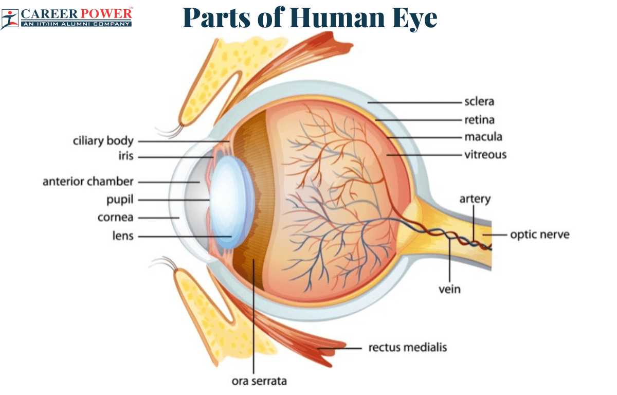
Vision-related issues can arise from various factors, affecting the ability to see clearly and perceive details. These conditions may stem from structural abnormalities, diseases, or aging processes, leading to challenges in visual clarity and focus. Understanding these conditions helps in identifying symptoms early, promoting better care and treatment options.
Refractive Errors
One of the most common vision problems occurs when light entering the visual system is not properly focused, leading to blurred or distorted images. Nearsightedness, farsightedness, and astigmatism are examples of refractive errors, where the shape of the cornea or lens deviates from the ideal, causing images to appear out of focus. Corrective lenses are often used to compensate for these imperfections, helping individuals achieve clearer vision.
Age-Related Conditions
As people age, certain conditions related to the gradual changes in the structure and function of the visual system become more prevalent. Cataracts, for instance, involve the clouding of the lens, leading to diminished clarity. Another age-related disorder, macular degeneration, affects the central part of the retina and can result in the loss of sharp, central vision. Early diagnosis and treatment can slow the progression of these conditions and improve the quality of life for those affected.